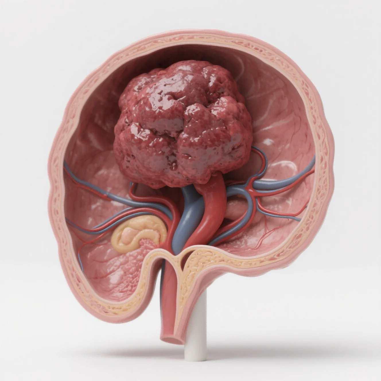
Move a tumor from the flank to its organ of origin and the experimental landscape changes. Blood supply, stromal signals, tissue pressure, local immune populations, and routes of spread begin to shape the response. An orthotopic breast tumor model is valuable because it exposes a candidate to that organ-specific setting, allowing predictive accuracy to be discussed in biological terms rather than as a generic promise.
That placement changes the question from whether a compound can slow a convenient mass to whether it can act within a more realistic tissue context. Vasculature, extracellular matrix, immune composition, oxygen, nutrients, and organ architecture can affect target dependence, drug penetration, invasion, and resistance.
Subcutaneous models remain valuable because tumors are accessible, measurement is straightforward, and studies can often be standardized efficiently. They exchange some tissue context for speed and visibility. Orthotopic designs accept greater technical and monitoring burden to test organ-specific behavior that a flank implant was never built to reveal.
A strong design starts by naming the feature the study needs to predict: primary growth, local invasion, metastatic spread, immune response, or organ-specific exposure. The team then selects and qualifies the model around that feature rather than around a general desire for greater complexity.
Tissue Context Changes Tumor Behavior
Tumor cells exchange signals with fibroblasts, endothelial cells, immune populations, and extracellular matrix. Those interactions can alter proliferation, angiogenesis, metabolism, and therapeutic sensitivity.
A mammary fat pad, liver, brain, lung, or another tissue-specific implantation site provides local cues that a flank implant may not reproduce. A tiered strategy may use both formats at different decision points within one program. In an orthotopic breast tumor model, the mammary environment can influence both local growth and treatment response.
Local oxygen and nutrient gradients can create regions with different growth rates and drug responses. Tissue pressure and vascular structure also affect compound delivery. A treatment that performs well against a uniformly accessible subcutaneous tumor may behave differently when exposure has to reach a deep and heterogeneous lesion.
Organ location can shape invasion and metastatic routes. Cells may enter local vessels, lymphatics, or adjacent structures in patterns linked to the primary site. Monitoring both the primary lesion and distant organs provides information about progression that a simple terminal tumor weight cannot capture. Feasibility criteria include the expected signal window and a plan for failed implantation.
These advantages come with tradeoffs. Surgery or image-guided implantation increases technical variability, and deep tumors are harder to measure. The gain in context is meaningful only when staff competency, model take, welfare monitoring, and endpoint sensitivity are sufficient to keep operational noise under control.
Imaging and Pathology Reveal Organ-Specific Response
Longitudinal imaging is central because deep lesions cannot usually be followed with calipers. Fluorescence or bioluminescence can track burden over time, while small-animal ultrasound or other modalities may suit particular organs.
The team standardizes signal calibration, acquisition settings, timing, and blinded analysis across groups. Spatially resolved sampling can show whether a biomarker change is confined to a particular compartment. Monitoring an organ-site tumor requires methods sensitive to deep and spatially irregular disease.
Imaging connects to anatomical and tissue evidence. H&E pathology can define lesion location, necrosis, and invasion; immunohistochemistry can assess proliferation, vascular or pathway markers; qPCR, ELISA, or related assays can examine molecular response. Concordant endpoints make an apparent imaging change easier to interpret.
Deep lesions place unusual demands on monitoring. Jennio Biotech provides orthotopic, metastatic, PDX, and humanized tumor-model approaches, with imaging, histopathology, immune profiling, and molecular readouts available according to the study design. It is useful to the study when those components share a calendar and sampling map; otherwise, platform breadth simply creates more disconnected observations.
Immune-cell subsets and cytokines may add another layer when the host system and therapy make them relevant. Sampling follows spatial differences, since blood, primary tumor, and metastatic sites can show different biology. A single pooled measurement may obscure the compartment in which treatment is acting.
Selecting the Right Orthotopic Design
Cell-derived designs support relatively standardized efficacy comparisons, whereas orthotopic PDX work may better preserve aspects of patient-tumor heterogeneity. Humanized models can address selected immune-therapy questions.
Combined primary-and-metastatic designs are useful when the product hypothesis depends on both local control and dissemination. Necropsy procedures include a consistent search for small or anatomically concealed lesions. Spatial lesion maps can then connect organ distribution with imaging sensitivity and treatment timing.
For an orthotopic breast tumor model, technical feasibility reaches beyond implantation success. Imaging sensitivity, primary-site sampling, metastatic surveillance, welfare limits, and tissue orientation all affect whether the organ-specific response can be interpreted at the planned dose and schedule.
Selection considers cell or tissue source, host immune status, implantation site, expected take rate, growth window, metastatic pattern, and available readouts. For breast cancer, a mammary-site model may suit primary-tumor biology, while a bone-destruction or bone-metastasis design is appropriate only when that mechanism drives the decision. Image acquisition follows the treatment and tissue-collection calendar.
The useful orthotopic study is the one whose added biological context answers a question that the flank model cannot. If organ placement changes invasion, exposure, metastatic spread, or immune response in a measurable way, the extra complexity has a purpose. If it does not, a simpler design may serve the program better.